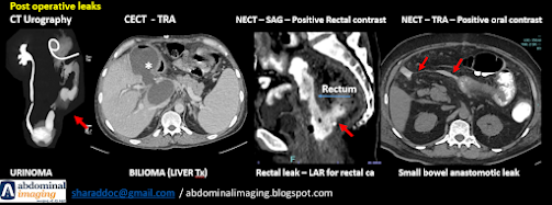
Postoperative
Abdominal Complications on CT: A Review
Introduction & Importance of CT
Postoperative
complications after abdominal surgery are common and require accurate
diagnosis.
Computed Tomography
(CT) is the primary imaging modality for evaluation [PubMed 37024236,
PubMed 19752834].
CT guides crucial
management decisions: conservative vs. interventional vs. surgical
treatment [PMC4956629].
Challenge:
Differentiating expected postoperative changes from true pathology is key
[PMC4330234].
General Considerations - Timing & Comparison
Timing is Crucial:
Early post-op:
Expect small fluid/gas collections; typically regress [PubMed 19752834].
Persistent/increasing
findings are concerning.
Free air (esp.
after laparoscopy) should decrease over time [PMC4330234].
Comparison Scans:
Essential to
compare with baseline/previous studies.
Helps track
evolution of findings [PMC4330234].
General Considerations
- CT Protocols
Modality: Multidetector CT (MDCT) preferred [PMC4330234].
Contrast:
IV Contrast: Venous phase often sufficient for
infection/collections [PMC4330234]. Arterial phase for active bleed/vascular
issues.
Delayed Phase: Essential for urinary leaks (CT Urography)
[PMC6176303, OUP Urinoma].
Oral/Rectal Contrast:
Selective use for bowel evaluation, suspected leaks [PMC7289697].
Complication: Hemorrhage & Hematoma
Clinical Clues: Early
post-op, hemodynamic instability, ↓Hb, drain output.
CT Findings:
Active Bleeding: Contrast extravasation ("blush")
[PMC4956629].
Hematoma: Collection,
often near surgical bed. Acutely hyperdense on non-contrast CT. Density
decreases over time.
Complication: Abscess / Infection
Clinical Clues:
Fever, ↑WBC, localized pain, purulent drainage (often 5-7+ days post-op)
[PMC4330234].
CT Findings:
Low-attenuation
fluid collection [PMC5510129].
Rim Enhancement:
Often thick, irregular (key feature) [ResearchGate Neck Abscess].
Gas bubbles /
Air-fluid level (highly suggestive) [PMC5510129].
Surrounding fat
stranding / inflammation [PMC5510129].
Differentiation from simple fluid/hematoma is critical.
Complication: Anastomotic Leak
Clinical Clues:
Fever, tachycardia, pain, ileus, change in drain output (feculent, urine,
bile), ↑WBC (often 5-10 days post-op) [PMC7289697].
CT Findings:
Extraluminal Gas: Near anastomosis,
more than expected/increasing [PMC7289697].
Extraluminal Contrast: Specific sign if
oral/rectal/delayed IV contrast used [PMC7289697].
Adjacent Fluid Collection/Abscess:
Common finding [PMC7289697].
Wall thickening /
Inflammation at anastomosis [PMC7289697].
Complication: Bowel Obstruction
Clinical Clues:
Nausea, vomiting, distension, cramping pain, obstipation.
CT Findings (Ileus):
CT Findings (Subacute obstruction):
CT Findings
(Mechanical Obstruction):
Dilated proximal
bowel (>3 cm SB) / Collapsed distal bowel [AJR Gastric Carcinoma Resection -
afferent loop].
Clear Transition
Point [AJR Gastric Carcinoma Resection - afferent loop].
Identify cause:
Adhesions (most common late cause), hernia, etc. [PubMed 19752834, PMC4729712].
Ileus: Diffuse dilatation, no transition point
(common early).
Complication: Bowel Obstruction - Worrisome Signs
Closed-Loop
Obstruction:
Bowel obstructed at two points.
High risk of
ischemia/strangulation [PMC4729712].
Look for
C/U-shaped loops, radial configuration, whirl sign.
Signs of Ischemia:
Bowel wall thickening.
Submucosal edema (Target sign).
Lack of mural enhancement.
Pneumatosis
intestinalis (intramural gas).
Portal venous gas.
Requires urgent
attention!
Complication: Urinoma / Urinary Leak
Clinical Clues:
Post GU/Gyn/Pelvic surgery. Flank/abdominal pain, mass, fever, drain output
(urine), ↑Creatinine [PMC6176303, OUP Urinoma].
CT Findings:
Peri-nephric or pelvic fluid collection [OUP
Urinoma].
Key: Extravasation of IV contrast into the
collection on DELAYED images [PMC6176303, OUP Urinoma].
May see associated
hydronephrosis.
Clinical Clues: Often
delayed presentation (weeks/months/years). Pain, infection, obstruction, fistula,
or incidental finding.
CT Findings:
Soft-tissue mass
[AJR Retained Sponges].
Spongiform /
Whorled pattern with trapped gas bubbles [AJR Retained Sponges, AJR Retained
Sponge Diagnosis].
Radiopaque Marker:
Diagnostic if present (check scout images!) [AJR Retained Sponges].
May have enhancing
capsule (granuloma) or associated abscess [AJR Retained Sponges, AJR Retained
Sponge Diagnosis].
Can mimic tumor,
abscess, hematoma.
Complication: Wound Issues
Clinical Clues:
Incisional pain, erythema, discharge, palpable fascial defect.
CT Findings:
Infection: Subcutaneous/intramuscular
fluid collections +/- gas near incision [ResearchGate CT diagnosis].
Hematoma: Collection within wall
layers.
Dehiscence: Fascial layer separation
+/- herniation [ResearchGate CT diagnosis, PubMed 19752834].
Conclusion
CT is Essential:
The cornerstone for diagnosing postoperative abdominal complications [PubMed
37024236].
Context Matters:
Integrate imaging findings with clinical picture and surgical history.
Know Normal vs.
Abnormal: Familiarity with expected post-op changes is vital [PMC4330234].
Comparison
Accurate Reporting:
Precise description and addressing the clinical question guides appropriate
management.
References
PubMed 37024236: Complications after abdominal surgery.
Radiologia (Engl Ed). 2023.
PubMed 19752834: [Postoperative imaging of the peritoneum
and abdominal wall]. J Radiol. 2009.
PMC4956629: Multidetector CT... ventral hernia repair.
Insights Imaging. 2016.
PMC4330234: Pictorial
review... laparoscopic appendectomy. Insights Imaging. 2015.
PMC6176303: Urinoma: Prompt Diagnosis and Treatment...
Cureus. 2018.
OUP Urinoma: Abdominal urinoma after oncologic
gynaecological surgery. Postgrad Med J. 2022.
PMC7289697: Normal and Abnormal Postoperative Imaging
Findings after Gastric... Surgery. Korean J Radiol. 2020.
ResearchGate Neck Abscess: CT differentiation of abscess and
non-infected fluid... Acta Radiol. 2013.
PMC5510129: CT findings... postoperative abdominal infection
patients with pancreatic carcinoma. Pak J Med Sci. 2017.
PMC4729712: Imaging the postoperative patient: long-term
complications... Insights Imaging. 2016.
AJR Gastric Carcinoma Resection: CT Findings... After
Gastric Carcinoma Resection. AJR Am J Roentgenol. 2002.
Radiopaedia SBO: Small bowel obstruction | Radiology
Reference Article | Radiopaedia.org (Summarizes findings from multiple
sources).
AJR Retained Sponges:
Imaging of Retained Surgical Sponges... AJR Am J Roentgenol. 2003.
AJR Retained Sponge Diagnosis: Retained surgical sponge:
diagnosis with CT and sonography. AJR Am J Roentgenol. 1988.
ResearchGate CT
diagnosis: CT diagnosis of postoperative abdominal complications. Abdom
Imaging. 2004.


Comments
Post a Comment