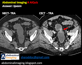
Differentiating Pathological stricture from Bowel spasm: Practical tips to solve the problem
Introduction:
- Small bowel spasm is a frequently seen finding in contrast MDCT. This can happen despite administration of Buscopan.
- It can mimic short / multiple stricture.
Problem solving:
1. Ensure optimal bowel distension
2. Compare with the plain scan
3. Use multiplanar reformats
4. Identification of similar areas of spasm - multiplicity
5. Jejunum always enhances more than ileus due to its fold density.
6. Collapsed bowel loops enhancement is more than distended bowel, which, further confound the problem. However, mucosal hyper enhancement is absent
7. Look for Inflammatory signs in the adjacent mesentry - Prominent vasa recta / fat stranding / adjacent lymphadenopathy
8. Length of involvement - long segment favors pathological stricture
9. Tight stricture will usually cause proximal dilatation and fecalization.
10. Degree and asymmetry of wall thickening. Asymmetry is more likely to be seen in pathological bowel.
Prepared by:
Dr. Reema
Dr. Sharad Maheshwari
6.7.2023

Comments
Post a Comment