Eosinophilic gastroenteritis: Pathophysiology & Imaging appearance
More common in pediatric population
Affects stomach and the small intestine
Pathological Hallmark:
Characterized by eosinophilic infiltration of the intestinal wall
3 sub types based on the infiltration of the wall: mucosal / muscular / serosal.
Mucosal type is the most common, which may progress to muscular & serosal layer (centrifugal disease)
Pathophysiology:
Concomitant allergic disorders and family history of Atopy is common and, suggests that it may be a result from "immune dysregulation" in response to allergic reaction
Eosinophils induce a significant inflammatory response
Diagnosis:
Symptoms (abdominal pain / diarrhoea / malabsorption), histology and exclusion of other causes
Treatment: Steroids
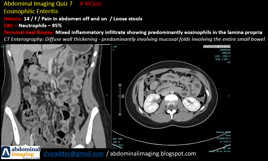
Imaging (CT Scan):
- Diffuse thickening of the mucosal folds / Intestinal wall thickening
- Ascites & Obstruction
- Halo Sign: Layering of the wall (submucosal edema)
- Araneid-limb-like (spider legs): orally administered oral contrast coats the mucosal folds and sinks into the sinuses between the folds. Similar to "accordion sign" seen in pseudomembranous colitis.
Non of the signs are specific
Other findings: strictures / polypoid lesions with ulcerations / thickened and flattened valvulae conniventis / mural thickening with luminal narrowing and rarely obstruction.
Prepared by Dr. Sharad Maheshwari
23.12.2022
Updated: 26.12.2022
References:
1. https://www.ncbi.nlm.nih.gov/pmc/articles/PMC5095570/
2. https://pubs.rsna.org/doi/epdf/10.1148/rg.220004
3. https://dirjournal.org/content/files/sayilar/42/buyuk/pdf_DIR_418.pdf
Comments
Post a Comment