CT LIVER DONOR REPORTING MADE EASY:
Key findings the surgeons want to know:
1. Liver Fat quantification:
a. Liver attenuation difference (LAI) on non-contrast CT scan, or
b. Proton density fat fraction (PDFF) calculation on MRI
Contrast Protocol:
1. Use higher iodine delivery per second (Flow rate of 4 to 5 cc / seconds and 370 or 400 mg iodine strength contrast)
2. At 64 slice scanner, post trigger: 6 seconds (early arterial) 20 seconds (portal phase) and 70 seconds (hepatic venous)
2. Hepatic Arterial Anatomy:
a. Aberrant origin of right hepatic artery (RHA) from superior mesenteric artery (SMA)
b. Aberrant/accessory left hepatic artery (LHA)
c. Diameter of RHA
d. Origin of segment 4 artery
e. Distance between the first right lobe hepatic branch of RHA after its origin. When the segment 4 artery arises from the RHA, give the distance from the segment 4 origin to the first right lobe hepatic branch.
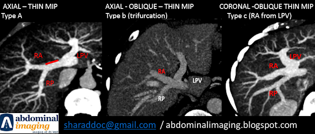
3. Portal Vein Anatomy:
a. Distance between the origin and branching of right portal vein (RPV) into anterior and posterior segments
b. Look for trifurcation and aberrant origin of the right anterior segment of the RPV from LPV - two veins for anastomosis
c. Any major cross over portal vein branch from left to the right or vice-versa
c. Types that reject the donor - Type D, Type E and single portal vein
4. Hepatic Vein Anatomy:
a. Accessory right hepatic vein (RHV) and its distance form the origin of RHV
b. Segment 5 tributaries and diameter that join the middle hepatic vein (MHV)
c. Segment 8 tributary and diameter - usually drains into the MHV
d. Segment 4 tributaries and where they join - usually segment 4b joins the MHV. Segment 4 a vein may join either the left hepatic vein (LHV) or MHV.
e. Presence of fissural veins draining into the left system. It may give a leverage to the surgeon to take MHV with the donor liver for harvest
5. Liver volumes:
After 3D model is made, do virtual resection, whereas possible, in the presence of the transplant team to increase the accuracy
a. Usually the resection is to the right of middle hepatic vein, cutting the segment 5 tributary to the MHV, followed by reconstruction.
b. Sometimes the resection is to the left of the middle hepatic vein with the sacrifice of the segment 4b vein.
b. Congestion volume calculation, if there is a decision to sacrifice a vein.
Prepared by: Dr. Sharad Maheshwari
Inputs from: Dr. Ruchi Rastogi and Dr. Sandeep Vohra
Prepared on: 6.10.2022
Updated: 11.10.2022


very nice and easy to understand
ReplyDeleteThank you
ReplyDelete