How to improve communication and highlight important findings in your report

1. If the referral note is lacking the right information, talk to the physician& understand their expectation from the scan
2. If the findings are complicated, it will be important to divide the body of report in two parts. For eg. in a pancreatic ca study, the first part could be details description of the tumor, its local extension and vascular anatomy. The second part of the report can describe rest of the findings.
3. Provide one or two relevant images, in the report itself, which are representative of the key finding. sometimes description can be long, of which the physician creates a visual impression, and may struggle to find the image of interest, from sea of images, provided in the 5 or 6 films.
4. Critical findings for. eg obstructing calculus causing acute ureteric obstruction are self explanatory in the image itself. It also covers any errors in the body of report.
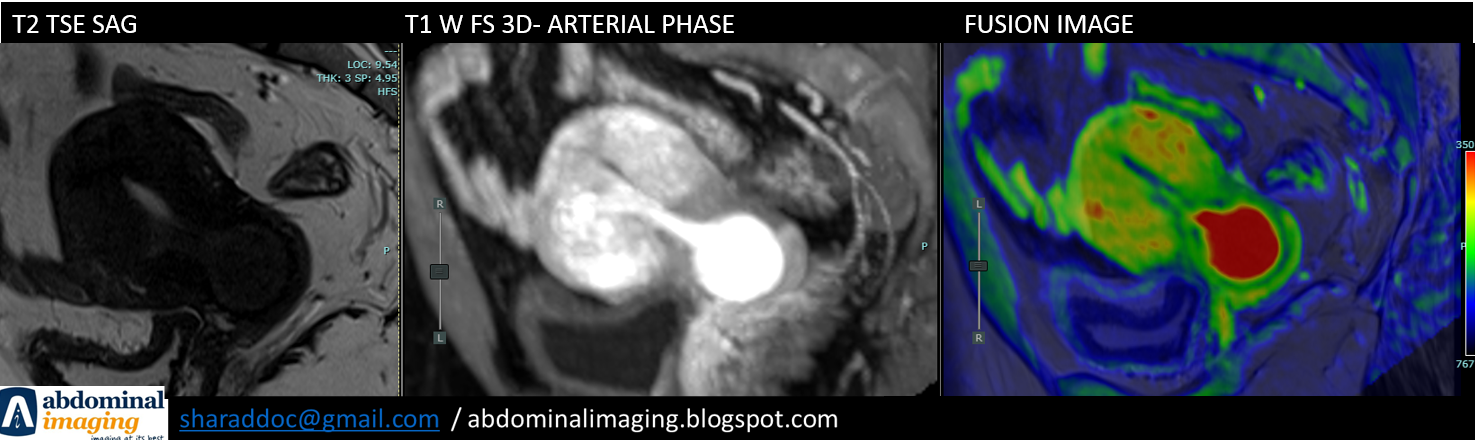
Case: Example below: Highly vascular polyp in the cervix, arising from the endometrium, that could lead to torrential bleeding, on just simple probing itself. Image is striking and gives the impact. Whereas, communication at the same time to gynecologist, changed the plan to either do embolization, before attempting polypectomy or plan a hysterectomy.
5. Impression should have point wise all relevant findings, which will help in the patient management. First point, should be always answer the specific question asked by the physician. Some of the findings, may not be directly related to current clinical problem may need a mention. For eg. incidental vertebral body compression fracture in a case of severe osteopenia of the spine, will eventually need medical attention.
6. Calling and discussing an important finding, which may not be possible to put in words. Also, the physicians don't like to read essays in their busy OPD or rounds.
7. Remember, every physician is not a specialist and may not be updated with your radiology lexicon
8. One of the best example could be a report of a "fistula study", where description can be a challenging and confuse the physician even before coming to a conclusion. It may be prudent to report in a defined fashion, describing each fistula separately with its ramifications, opening, abscess etc. Give relevant finding on key images and even drawing on a sheet, that can be attached.
9. Any limitations, like patient motion, artifacts from implants or very obese patient reduces scan resolution etc should be mentioned
10. Whenever possible give a final diagnosis with possible differential. eg. classical hemangioma should be a single and final diagnosis. While FNH vs Adenoma can be differential.
11. Avoid long sentences, when there are multiple findings in the same region of interest. You can break and divide the findings in two separate sentences.
12. It may be good idea to describe the body of report as multiple stations. For eg. Adrenal and the urinary system as one paragraph. Hepatobiliary and pancreas as one paragraph etc
13. Satisfaction of search: It is an interesting phenomenon, where your interest in rest of the findings is lost, as you find the main abnormality based on the history. It will be prudent to see every scan in a defined pattern, regardless of the specific question raised. For eg. If a scan is done for appendicitis, don't start with looking at the appendix. Follow the pattern you have created, which could start from description of the liver etc. Appendix you will see, when its turn comes in your pattern. Seems complicated, but very simple after everyday use.
14. All the normal organs can be clubbed in a one sentence or two. For eg: The liver, spleen, pancreas, gallbladder, intra and extra hepatic bile ducts are unremarkable.
15. Comparison from the previous scans is vital, especially in oncology.
16. In the end of report you could mention that the case was discussed with Dr. XYZ and remind them to read the body of report
17. If you obtain an opinion from your colleague, with their consent, you can mention their name
18. When you are unable to give explanation or significance of an imaging finding, its is always a good idea to mention that you don't know and ask for more time to work on it.
19. Avoid terms like advised or recommended. Instead use: suggested / if of concern
20. Clinical correlation vs specific inputs: Based on your evidence based practice, you can provide specific suggestions. Some of the referring physicians don't like to be told what to be done, in that case you may not write.
Below is an example of a Impression given by me in a case of ovarian ca, which came for first follow up after surgery:
"Impression:
- Prominent vaginal cuff on the left side with heterogeneous enhancement as
described.
- Subtle nodular thickening is seen in the fat deep to the right abdominal wall.
The significance is undetermined. Clinical correlation with CA-125,
and short follow-up is suggested.
- No evidence of ascites or significant lymphadenopathy.
- Hepatomegaly and splenomegaly with underlying changes early changes of
portal hypertension. Liver parenchymal disease to be ruled out.
Further gastroenterology consultation is suggested."
Prepared by: Dr. Sharad Maheshwari
Published: 27.9.2022
Updated: 28.12.2022
good
ReplyDeleteThank you
DeleteExcellently decoded
ReplyDeletethank you
Deletegreat details of Normal findings should be avoided as also un necessary details
ReplyDeleteYes! All the normal organs can be clubbed in a sentence. For eg: The liver, spleen, pancreas, gallbladder, intra and extra hepatic bile ducts are unremarkable.
DeleteGood
ReplyDeletethank you
DeleteFor fistulogram studies I actually draw the findings in the report for clinicians to understand
ReplyDeleteThanks
DeleteWell summarized including so many good pointers to remember. Great Job, Sharad!
ReplyDeleteThank you
ReplyDelete