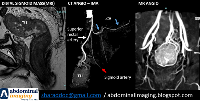
Anatomy of Inferior mesenteric artery(IMA) is important while evaluating for rectal and distal sigmoid ca on cross sectional imaging
Anatomy: Branches of IMA:
1. Left colic artery
2. Sigmoid artery
3. Superior rectal artery
Anatomical variation:
1. Early origin of LCA
2. Common trunk of LCA and sigmoid artery
3. Trifurcation of LCA, sigmoid artery and the superior rectal artery
Surgeons approach during laparoscopic radical resection:
1. en-block lymph node resection at the origin of IMA
2. Tie the IMA:
- High tie at the artery at the origin. Compromises LCA supply to the
descending colon and increase risk of anastomotic leak.
- Low tie: preserves the LCA
Win-Win situation:
Early and separate branch of the LCA
Case discussion:
- 47 / M - high rectum-distal sigmoid mass
- T3
- Sphincter spared
- Tumor primarily supplied by superior rectal artery
Management Plan: Laparoscopic radical resection
Anatomical consideration: Spare the LCA as it originates separately
Further reading:
https://www.ncbi.nlm.nih.gov/pmc/articles/PMC6113723/

Comments
Post a Comment